Background
Pneumoconiosis is the general term for lung disease caused by inhalation and deposition of mineral dust.Pneumoconiosis caused by asbestos inhalation is called asbestosis. The word asbestos is derived from Greek and means inextinguishable, and asbestos is a group of naturally occurring, heat-resistant fibrous silicates. Asbestos fibers are long and thin (length-to-diameter ratio >3) and may be either curved or straight. The curved fibers are called serpentine (chrysotile is the prime example), and the straight fibers are amphiboles. Several different types of amphiboles (ie, amosite, anthophyllite, tremolite, actinolite, crocidolite) have been recognized. Chrysotile is by far the most common type of asbestos fiber produced in the world and accounts for virtually all asbestos used commercially in the United States.
Production and use of asbestos increased greatly between 1877 and 1967. In the 1930s and 1940s, scientists recognized a causal link between asbestos exposure and asbestosis. In the 1950s and 1960s, researchers established asbestos as a predisposing factor for bronchogenic carcinoma and malignant mesothelioma.
Note the image below.
 Asbestos pleural plaques.
 Causes
In modern times, the risk to persons in the United States occurs mainly through the processing, manufacturing, and end-use of asbestos.
Manufacturers commonly use asbestos in the following products:
Manufacturers commonly use asbestos in the following products:
- Products containing asbestos cement - Pipes, shingles, clapboards, sheets
- Vinyl-asbestos floor tiles
- Asbestos paper in filtering and insulating products
- Material in brake linings and clutch facings
- Textile products - Yarn, felt, tape, cord, rope
- Spray products used for acoustical, thermal, and fireproofing purposes
- Insulation workers
- Boilermakers
- Pipefitters
- Plumbers
- Steamfitters
- Welders
- JanitorsImaging
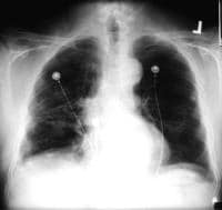
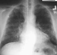 Case 1. Postero-anterior (PA) chest radiograph in a 58-year-old man with a history of occupational exposure to asbestos shows right diaphragmatic pleural plaque calcifications, linear calcification along the left pericardium, and bilateral pleural plaques along upper ribs.
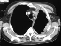 Case 4. The soft-tissue window setting of this chest computed tomography (CT) scan shows the envelope-like mass along the pleural surface surrounding the lung. This was a mesothelioma.
Pleural calcification
On chest radiographs, the prevalence of calcification in pleural plaques is reported to be 10-15%. In profile, calcified plaques appear as opaque lines that lie parallel to the chest wall, mediastinum, pericardium, and diaphragm. Viewed en face, calcified plaques are seen as irregular, heterogeneous densities, the so-called holly leaf. The presence of bilateral, superior diaphragmatic surface calcifications with clear costophrenic angles is virtually pathognomonic for asbestos-related pleural disease. (See the image below.)
Case 1. Postero-anterior (PA) chest radiograph in a 58-year-old man with a history of occupational exposure to asbestos shows right diaphragmatic pleural plaque calcifications, linear calcification along the left pericardium, and bilateral pleural plaques along upper ribs.