Thursday, August 4, 2011

Chance Fracture

Chance Fracture of the Spine


- Discussion:
    - Chance frx & posterior ligament rupture (variant of flexion distraction injury pattern) may
          present w/ minor anterior vertebral compression;
          - in Chance frx, the anterior column fails in tension (along w/ the middle and posterior columns),
                 where as flexion distraction fracture involve compression of the anterior column and
                 distraction of the middle and posterior columns;
    - approx 1/2 of pts w/ flexion distraction injury pattern have primarily ligamentous rupture;
          - rupture usually includes interspinous ligament, ligamentum flavum, facet capsule, posterior
                 annulus, and thoracodorsal fascia;
    - whether the injury is purely ligamentous or includes a fracture thru vertebral body, all three columns
          rupture in distraction (tension);
    - often these are misdiagnosed as a compression frx;
          - the occurance of a traumatic compression fracture in a young patient (following MVA) should
                 raise the possibility of a Chance fracture;
          - either good quality AP view is necessary to rule out posterior element injury, or a CT scan is
                 required (if the AP view remains equivocal);

- Exam:
    - seldom assoc w/ neurologic compromise unless
    - abdominal injuries are common and occur in upto 50-60% of patients;
    - references:
          - The epidemiology of seatbelt associated injuries.
                 PA Anderson et al.  J. Trauma. Vol 31. 1991. p 60-67.

- Radiographs:
    - significant translation on lateral;
    - anterior wedging may be minimal;
    - often only a portion of the vertebral body will be involved (half ligamentous and half bony injury);
    - look for frx line extending thru spinous process, lamina, pedicles, & portion of the vertebral body;
    - often the AP view will best show the posterior element injury (lamina frx will appear as a "lazy W")  

- CT Scan: is often ordered to help make the diagnosis;


- Non Operative Treatment:
    - Chance Frx may initially be unstable, but after 2 weeks there will be sufficient bony healing
           to allow fitting for an orthosis;
           - patients w/ partial vertebral body involvement (half bony injury and half ligamentous injury)
                  may be candidates for non operative treatment is alignment is acceptable;
    - candidates for non operative treatment should have less than 15 deg of kyphosis;
    - patients should be fitted for a custom molded hyperextension orthosis;
    - fractures below L3 may require the addition of a thigh extension;

- Indications for Operative Treatment:
    - w/ Ligamentous Chance Injury, soft tissue healing is unreliable, and about
           half of all patients treated non operatively will have poor outcomes;
           - progressive kyphosis is one of the major complications w/ non-op Rx;


Thursday, July 14, 2011

Afferent Pupillary Defect

Marcus Gunn pupil (relative afferent pupillary defect)

is a medical sign observed during the swinging-flashlight test[1] whereupon the patient's pupils constrict less (therefore appearing to dilate) when a bright light is swung from the unaffected eye to the affected eye.

The affected eye still senses the light and produces pupillary sphincter constriction to some degree, albeit reduced.

The most common cause of Marcus Gunn pupil is a lesion of the optic nerve (proximal to the optic chiasm) or severe retinal disease. It is named after Scottish ophthalmologist Robert Marcus Gunn.[2]


  Look for an afferent pupillary defect (RAPD - also known as Marcus Gunn Pupil) by:

a.       The Swinging Flash Light Test
b.      The pupils must react to light in order to perform the test
c.       Not a test of the pupil per se—but we do the test when we look at the pupil
d.      Tests the optic nerve function
e.       Relies on a difference between the two optic nerves—one must be different from each other


Wednesday, July 6, 2011

Subperiosteal Abscess


Bilateral ethmoidal sinusitis with subperiosteal abscess of the left medial wall.

amaurosis fugax

Amaurosis fugax (Latin fugax meaning fleeting, Greek amaurosis meaning darkening, dark, or obscure) is a transient monocular visual loss.

The experience of amaurosis fugax is classically described as a transient monocular vision loss that appears as a "curtain coming down vertically into the field of vision in one eye;" however, this altitudinal visual loss is relatively uncommon. In one study, only 23.8 percent of patients with transient monocular vision loss experienced the classic "curtain" or "shade" descending over their vision.[43]

Other descriptions of this experience include a monocular blindness, dimming, fogging, or blurring.[44] Total or sectorial vision loss typically lasts only a few seconds, but may last minutes or even hours. Duration depends on the etiology of the vision loss. Obscured vision due to papilledema may last only seconds, while a severely atherosclerotic carotid artery may be associated with a duration of one to ten minutes.[45] Certainly, additional symptoms may be present with the amaurosis fugax, and those findings will depend on the etiology of the transient monocular vision loss.

MANY CAUSES OF AMAUROSIS FUGAX

Cell and Flare

Cell and Flare


Physical exam finding often associated with acute iritis

Tuesday, June 28, 2011

Resuscutation Sequence: Unwitnessed VFib Arrest

1.  2 minutes of CPR (prime the pump)
2.  Shock (120-200 J biphasic defibrillator)
3.  RESUME CHEST COMPRESSIONS immediately following shock (to eliminate delays)
4.  Check pulse (10 seconds during second round of chest compressions)
5.  If no pulse: Shock 200 J
6.  RESUME CHEST COMPRESSIONS immediately following shock

Friday, June 24, 2011

Local Anesthesia Toxicity (Lidocaine)

Local Anesthetic Agents Used Commonly for Infiltrative Injection
AgentDuration of ActionMaximum Dosage Guidelines (Total Cumulative Infiltrative Injection Dose per Procedure*)


Amides

Lidocaine (Xylocaine)Medium (30-60 min)Without epinephrine: 4.5 mg/kg; not to exceed 300 mg
Lidocaine with epinephrineLong (120-360 min)With epinephrine: 7 mg/kg


Bupivacaine (Marcaine)Long (120-240 min)Without epinephrine: 2.5 mg/kg; not to exceed 175 mg total dose
Bupivacaine with epinephrineLong (180-420 min)With epinephrine: Not to exceed 225 mg total dose


Bottom line Lidocaine 1% => 10mg per ml = Don't give more then 30 mls


Thursday, June 23, 2011

Pediatic Resuscitation: SVT

Stable SVT

Vagal maneuvers: 
  1. Ice to face
  2. Blow through straw
Adenosine:
  1. Initial: 0.1 mg/kg  (max 6 mg)
  2. Susequant: 0.2 mg/kg (max 12 mg)
Consider alternative medication:
  1. Amiodarone: 5 mg/kg (over 20-60 min)
  2. Procainamide: 15 mg/kg (over 30-60 min)
_____________________________________________
Verapamil should not be used as blocking agent for in infants...Bradycardia and Hypotension


________________________________________


Unstable SVT:

Cardioversion:
  1. First dose: 0.5 - 1 J/kg
  2. Second dose: 2J/Kg

Monday, June 20, 2011

Scarlet Fever

CAUSED BY: Group A Strep


Sand paper rash:


Pastia's Lines


Strawberry Tongue


Sunday, June 19, 2011

Kawasaki's

My HEART

Mucosal involvement (strawberry tongue/fissured lips)
Hands (erythema palms/soles)
Eyes (conjuctivtis)
Adenopathy (cervical)
Rash: (usually trunk)
Temp: > 5 days

Criteria for Dx is Temp for 5 days and any 4 of the above

------------------------------------

Additional Mnemonic: CRASH and Burn


C-Conjunctivitis
R-Rash
A-Adenopathy
S-Strawberry tongue
H-Hand and feet swelling

Burn-fever for more than 5 days

Saturday, June 18, 2011

Pediatric Fever without focus Management

Terrible T's: cyanoisis

 Tetrology of Falot

a congenital heart defect which is classically understood to involve four anatomical abnormalities (although only three of them are always present). It is the most common cyanotic heart defect, and the most common cause of blue baby syndrome.


Tricuspid Atresia

Tricuspid fails to form (unopened).  No blood flow from RA to RV.  Tiny RV.  Blood instead shunts across Foramen Ovale or ASD into Pulmonary Arterial system for oxygenation.

Friday, June 17, 2011

Salter Harris

SALTER mnemonic for classification

The mnemonic "SALTR" can be used to help remember the first five types.  This mnemonic requires the reader to imagine the bones as long bones, with the epiphyses at the base.
  • I - S = Same (or Straight across). Fracture of the cartilage of the physis (growth plate)
  • II - A = Above. The fracture lies above the physis, METAPHYSIS. (MOST FREQUENT 75%)
  • III - L = Lower. The fracture is below the physis in the epiphysis.
  • IV - T = Through. The fracture is through the metaphysis, physis, and epiphysis.
  • V - R = Rammed (crushed). The physis has been crushed. (May dx on mechanism alone)

Thursday, June 16, 2011

Pediatric Fever

Historically, children aged 3 months to 3 years with rectal temperatures of 38.5o C (101.3 F) or higher had a risk of 2-4% for occult bacteremia.[1, 2] The leading cause of bloodstream infection was S pneumoniae, followed by H influenzae type b. With the introduction of effective vaccines for these pathogens, the incidence and epidemiology of childhood bacteremia in the immunologically normal host has changed.
The incidence of occult bacteremia in this population now ranges from 0.5-1%; moreover, 2 out of every 3 blood isolates from these children represent an artifact (contamination) and not a true pathogen.[3]S pneumoniae and E coli are the most common pathogens, accounting for two thirds of cases. In infants with S pneumoniae, many isolates are strains not covered by the currently available heptavalent conjugate vaccine.

Who do we need to get cath urine on???

Urine Cath Specimen: (from Boston Criteria for fever without focus)

< 6 month old males circumcised
<12 month old males uncircumcised
<24 month old females

Saturday, June 4, 2011

Oxygen Dissociation Curve

http://en.wikipedia.org/wiki/Oxygen%E2%80%93haemoglobin_dissociation_curve

The causes of shift to right can be remembered using the mnemonic, "CADET, face Right!" for CO2, Acid, 2,3-DPG, Exercise and Temperature.[1] Factors that move the oxygen dissociation curve to the right are those physiological states where tissues need more oxygen. For example during exercise, muscles have a higher metabolic rate, and consequently need more oxygen, produce more carbon dioxide and lactic acid, and their temperature rises.

--------------------------------------------

CADET, face Right!

  • CO2  
  • Acid
  • 2,3-DPG(aka 2,3-BPG)
  • Exercise
  • Temperature
Oxygen-haemoglobin dissociation curve

Friday, June 3, 2011

Asbestosis

Background

Pneumoconiosis is the general term for lung disease caused by inhalation and deposition of mineral dust.

Pneumoconiosis caused by asbestos inhalation is called asbestosis. The word asbestos is derived from Greek and means inextinguishable, and asbestos is a group of naturally occurring, heat-resistant fibrous silicates. Asbestos fibers are long and thin (length-to-diameter ratio >3) and may be either curved or straight. The curved fibers are called serpentine (chrysotile is the prime example), and the straight fibers are amphiboles. Several different types of amphiboles (ie, amosite, anthophyllite, tremolite, actinolite, crocidolite) have been recognized. Chrysotile is by far the most common type of asbestos fiber produced in the world and accounts for virtually all asbestos used commercially in the United States.
Production and use of asbestos increased greatly between 1877 and 1967. In the 1930s and 1940s, scientists recognized a causal link between asbestos exposure and asbestosis. In the 1950s and 1960s, researchers established asbestos as a predisposing factor for bronchogenic carcinoma and malignant mesothelioma.

Note the image below.
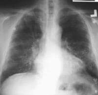
Asbestos pleural plaques. Asbestos pleural plaques. 

Causes

In modern times, the risk to persons in the United States occurs mainly through the processing, manufacturing, and end-use of asbestos.
Manufacturers commonly use asbestos in the following products:
  • Products containing asbestos cement - Pipes, shingles, clapboards, sheets
  • Vinyl-asbestos floor tiles
  • Asbestos paper in filtering and insulating products
  • Material in brake linings and clutch facings
  • Textile products - Yarn, felt, tape, cord, rope
  • Spray products used for acoustical, thermal, and fireproofing purposes
Examples of occupations associated with asbestosis include the following:
  • Insulation workers
  • Boilermakers
  • Pipefitters
  • Plumbers
  • Steamfitters
  • Welders
  • Janitors
     Imaging

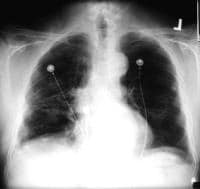
    Case 1. Postero-anterior (PA) chest radiograph in Case 1. Postero-anterior (PA) chest radiograph in a 58-year-old man with a history of occupational exposure to asbestos shows right diaphragmatic pleural plaque calcifications, linear calcification along the left pericardium, and bilateral pleural plaques along upper ribs. 

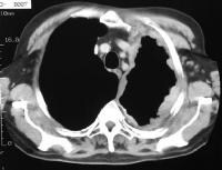
     Case 4. The soft-tissue window setting of this cheCase 4. The soft-tissue window setting of this chest computed tomography (CT) scan shows the envelope-like mass along the pleural surface surrounding the lung. This was a mesothelioma.

    Pleural calcification

    On chest radiographs, the prevalence of calcification in pleural plaques is reported to be 10-15%. In profile, calcified plaques appear as opaque lines that lie parallel to the chest wall, mediastinum, pericardium, and diaphragm. Viewed en face, calcified plaques are seen as irregular, heterogeneous densities, the so-called holly leaf. The presence of bilateral, superior diaphragmatic surface calcifications with clear costophrenic angles is virtually pathognomonic for asbestos-related pleural disease. (See the image below.)
    Case 1. Postero-anterior (PA) chest radiograph in Case 1. Postero-anterior (PA) chest radiograph in a 58-year-old man with a history of occupational exposure to asbestos shows right diaphragmatic pleural plaque calcifications, linear calcification along the left pericardium, and bilateral pleural plaques along upper ribs.


A-a gradient

Alveolar-arterial gradient

Pathophysiology sample values
BMP/ELECTROLYTES:
Na+=140 Cl=100 BUN=20 /


Glu=150
K+=4 CO2=22 PCr=1.0 \
ARTERIAL BLOOD GAS:
HCO3-=24 paCO2=40 paO2=95 pH=7.40
ALVEOLAR GAS:

pACO2=36 pAO2=105 A-a g=10
OTHER:
Ca=9.5 Mg2+=2.0 PO4=1
CK=55 BE=−0.36 AG=16
SERUM OSMOLARITY/RENAL:
PMO = 300 PCO=295 POG=5 BUN:Cr=20
URINALYSIS:
UNa+=80 UCl=100 UAG=5 FENa=0.95
UK+=25 USG=1.01 UCr=60 UO=800
PROTEIN/GI/LIVER FUNCTION TESTS:
LDH=100 TP=7.6 AST=25 TBIL=0.7
ALP=71 Alb=4.0 ALT=40 BC=0.5


AST/ALT=0.6 BU=0.2
AF alb=3.0 SAAG=1.0
SOG=60
CSF:
CSF alb=30 CSF glu=60 CSF/S alb=7.5 CSF/S glu=0.4
The Alveolar-arterial gradient (A-a gradient), is a measure of the difference between the alveolar concentration of oxygen and the arterial concentration of oxygen. It is used in diagnosing the source of hypoxemia.[1]

Equation

A-a gradient = PAO2 − PaO2[2]

On Room air ( 21 % ) and at sea level, a simplified version of the equation is:
Aa Gradient = (150 - 1.2*(PCO2)) - PaO2

Values and meaning

The A-a gradient is useful in determining the source of hypoxemia. The measurement helps isolate the location of the problem as either intrapulmonary (within the lungs) or extrapulmonary (somewhere else in the body).
A normal A-a gradient for a young adult non-smoker breathing air, is between 5-10 mmHg. Normally, the A-a gradient increases with age. For every decade a person has lived, their A-a gradient is expected to increase by 1 mmHg.
An abnormally increased A-a gradient suggests a defect in diffusion, V/Q (ventilation/perfusion ratio) mismatch, or right-to-left shunt.[3]
Because A-a gradient is approximated as: (150 - 5/4(PCO2)) - PaO2, the direct mathematical cause of a large value is that the blood has a low PO2, a low PCO2, or both. CO2 is very easily exchanged in the lungs and low PCO2 directly correlates with high minute ventilation; therefore a low arterial PCO2 indicates that extra respiratory effort being used to oxygenate the blood. A low PO2 indicates that at the patient's current minute ventilation (whether high or normal) is not enough to allow adequate oxygen diffusion into the blood. Therefore the A-a gradient essentially demonstrates a high respiratory effort (low arterial PCO2) relative to the achieved level of oxygenation (arterial PO2). A high A-a gradient could indicate a patient breathing hard to achieve normal oxygenation, a patient breathing normally and attaining low oxygenation, or a patient breathing hard and still failing to achieve normal oxygenation.
If lack of oxygenation is proportional to low respiratory effort, then the A-a gradient is not increased; a healthy person who hypoventillates would have hypoxia, but a normal A-a gradient


Wednesday, May 18, 2011

Cyanosis: Answer and Discussion

Answer:


3. Raynaud's phenomenon


Discussion:


Cyanosis is divided into two main types:

Central (around the core and lips) 

Peripheral (only the extremities are affected).

Cyanosis can occur in the fingers, including underneath the fingernails, as well as other extremities (called peripheral cyanosis), or in the lips and tongue (central cyanosis).

Central cyanosis


Central cyanosis is often due to a circulatory or ventilatory problem that leads to poor blood oxygenation in the lungs. It develops when arterial saturation of blood with oxygen is ≤85%. Cyanosis may not be detected until saturation is 75% in dark-skinned individuals.
Acute cyanosis can be a result of asphyxiation or choking, and is one of the surest signs that respiration is being blocked.

Causes

1. Central Nervous System:
2. Respiratory System:
3.Cardiac Disorders:
4.Blood:
5.Others:


Peripheral cyanosis


Peripheral cyanosis is the blue tint in fingers or extremities, due to inadequate circulation. The blood reaching the extremities is not oxygen rich and when viewed through the skin a combination of factors can lead to the appearance of a blue color. All factors contributing to central cyanosis can also cause peripheral symptoms to appear, however peripheral cyanosis can be observed without there being heart or lung failures. Small blood vessels may be restricted and can be treated by increasing the normal oxygenation level of the blood.

Causes

----------------

Pearls:


On Physical Exam cyanosis is best (most reliably) observed by looking at the tongue

Pulse-Oximetry cannot differentiate Methemoglobinemia

-----------------


Interesting Story:


The Fugates, a family that lived in the hills of Kentucky, are the most famous example of this hereditary genetic condition. Known as the Blue Fugates, Martin Fugate, settled near Hazard, Kentucky, circa 1800. His wife was a carrier of the recessive methemoglobinemia (met-H) gene, as was a nearby clan with whom the Fugates intermarried. As a result, many descendants of the Fugates were born with met-H.[7][8]

The 'blue men of Lurgan' were a pair of Lurgan men suffering from what was described as 'familial idiopathic methaemoglobinaemia' who were treated by Dr. James Deeny in 1942. Deeny, who would later become the Chief Medical Officer of the Republic of Ireland, prescribed a course of ascorbic acid and sodium bicarbonate. In case one, by the eighth day of treatment there was a marked change in appearance and by the twelfth day of treatment the patient's complexion was normal. In case two, the patient's complexion reached normality over a month-long duration of treatment.[9]

Question: Cyanosis

Question: Which is a cause of peripheral cyanosis?

1.  High Altitude
2.  Pulmonary Embolism
3.  Raynaud's Phennomenon
4.  Methemaglobinemia
5.  CHF

Friday, May 13, 2011

EM Board Review

Welcome to the EM Board review blog!!!   This blog is designed to assist anyone who is interested in Emergency Medicine, and particularly those who will be taking the Emergency Medicine Boards.  Each blog will illustrate a board review topic.  I hope to include articles, images, opinions...anything to make the topic more interesting and educational. The key is SHORT AND SWEET.

Here is a list topics that will be represented on the EM boards:

Trauma
Cardiovascular
Abdominal/GI
Thoracic/Respiratory
Pediatrics
Procedures/Sklls
Head, Ear, Eye, Nose, and Throat
ID
Neuro
OB/GYN
Heme/Onc
Toxicology
Endocrine/Metabolic
Environmental
Musculoskeletal
Psych
Renal/Uro
Derm
Prehospital/EMS

Week 1 starts with Thoracic/Respiratory because that's the point I'm at in my studying.  I'll plan to cycle back and include topics from the top of the list as I work my way through.

Hope you enjoy.